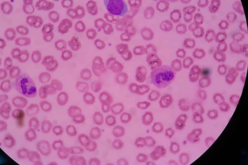
Live Cell Microscopy

Live cell microscopy allows practitioner to view specific details and provides the patient with a live snap shot of the condition of their blood. With this technique one drop of blood from the finger is placed on a microscope slide and is then viewed at a magnification 1500 times or more under a specialized high power darkfield microscope. The magnified blood sample is displayed on a video monitor and is visible to both the practitioner and client. Things that could be indicated by this method are:
– Altered PH
– Sign of free radical
– Certain vitamin or mineral deficiencies
– Digestion of protein and fat
– Sign of heavy metals
and much more.